扭到腳踝,能走路就算好了嗎?
踝關節扭傷 10 個常見問題:從剛扭到怎麼處理,到什麼時候可以回場上
扭到腳踝,冰敷幾天、腫消了、能走路,很多人就當作好了。
但腳踝外側扭傷是最常見的運動傷害,大約每 10 個人就有 4 個,之後會變成慢性踝關節不穩定(Chronic Ankle Instability, CAI),也就是一扭再扭1。
這篇用大家最常問的 10 個問題,依照研究整理成看得懂、做得到的答案。
先講結論
1. 扭到腳踝,需要照 X 光嗎?
不是每次扭傷都要照。醫師常用「Ottawa 足踝準則」來判斷。
有下面任一種情況,就應該就醫,評估要不要照 X 光41:
- 腳踝兩側突出的骨頭(外踝、內踝),後緣或尖端一壓就痛。
- 腳背外側的骨頭(第五蹠骨底部)或足弓內側中段的骨頭(舟狀骨)一壓就痛。
- 受傷當下和到醫院時,都沒辦法自己走 4 步。
研究顯示,這套準則幾乎不會漏掉骨折,還能減少 30% 到 40% 不必要的 X 光40。完整條文見文末補充。
另外,韌帶傷得多嚴重,受傷後 4 到 7 天再檢查會比較準216。如果恢復得不如預期,要回去就醫,醫師可能安排 MRI,排除骨頭或肌腱的其他傷害16。
2. 剛扭到,要冰敷嗎?要固定多久?
重點不在冰不冰,而是「先保護,再及早動起來」。
- 先保護:用護踝等外部支撐約 5 到 10 天16。韌帶有斷裂的人,用貼紮或護踝搭配運動治療,效果最好2。
- 及早動起來:有人指導的運動治療,比被動治療(由別人對你做、你自己不用出力的治療)好,因為它能幫腳踝找回穩定2。
- 冰敷:常見的軟組織處理原則 PEACE & LOVE 不鼓勵冰敷3;但目前還沒有高品質研究評估冰敷在其中的效果,正在進行的隨機試驗就是在比較這件事17。也就是說,冰敷好或不好,目前證據還不夠。
- 接下來:改用半硬式護踝等支撐,搭配 8 到 12 週的平衡與控制訓練(神經肌肉復健)16。
3. 可以吃止痛藥或消炎藥嗎?
可以,但要知道取捨。
用藥請依醫師或藥師的指示。
4. 腫消了、能走路,就代表好了嗎?
不一定。能走路,只代表日常生活大致沒問題。
約四成扭傷的人會變成慢性踝關節不穩定1;一項追蹤 474 人的研究,兩年後的比例是 34.4%4。
放著不管,長期會怎樣?
- 約三分之一合併軟骨受傷:統合 12 篇研究、2170 個慢性不穩定的腳踝,32.2% 有軟骨或骨軟骨傷害,大多在距骨(腳踝關節裡的一塊骨頭)19。
- 腳踝退化多半來自受傷:腳踝很少「自己」退化,通常是受傷後才出現;反覆扭傷造成的不穩定也可能導致退化20。
- 青少年扭傷也有長期影響:青少年運動時扭傷,3 到 15 年後追蹤,和沒受傷的人相比,症狀較多、功能與生活品質較差、平衡也較差21。另一項 10 人的小型研究,在 MRI 上看到傷側骨頭有退化跡象22。
5. 為什麼有人一扭再扭?哪些人比較容易?
同樣是扭傷,有些因素會讓變成慢性不穩定的機會變高。
一項追蹤 474 位第一次扭傷病人的多中心研究,找到三個獨立的危險因子4:
- 年紀:年紀愈大,風險愈高。
- 距骨軟骨受傷:MRI 上看到腳踝關節裡的軟骨有傷。
- 距舟韌帶受傷:連接距骨和舟狀骨的韌帶也受傷。
這是回溯性研究,可以幫助及早找出高風險的人,但不是每次扭傷都需要照 MRI4。
第一次扭傷就比較嚴重(第二級)的人,也比較容易再扭傷;不過扭傷分級本身準不準有疑慮,這個結果要保守看待18。
專業補充:危險因子的勝算比
| 預測因子 | 勝算比(OR) |
|---|---|
| 年齡(每增加一歲) | 1.07 |
| 距骨骨軟骨病灶(TOL) | 1.35 |
| 距舟韌帶(TNL)損傷 | 1.48 |
某些組合會讓風險加乘,例如距骨骨軟骨病灶合併男性或嚴重跟腓韌帶損傷,協同指數 3.24 到 4.454。這是回溯性研究,可以幫助及早辨識高風險的人,但不是每個扭傷都需要照 MRI。
6. 已經一扭再扭了,該怎麼練?
沒有一種方法能包辦全部,證據支持的是「組合」。
- 平衡+肌力訓練,再加徒手治療或乾針:比較 44 篇隨機對照試驗的研究發現,這個組合在功能、平衡和疼痛上效果排名最前5。
- 練多久:運動治療做超過 4 週,日常功能和平衡測驗都有明顯進步23。
- 別只練腳踝:加做髖部(臀部周圍)肌力訓練,平衡測驗和功能也會跟著進步6。
- 只練肌力不夠:只做彈力帶等肌力訓練 6 週,肌力和穩定感有進步,但跳躍和平衡測驗沒有改善27,所以要搭配平衡與功能性訓練。
- 想加點變化:用體感遊戲或虛擬實境練平衡,效果和傳統平衡訓練差不多,可以依喜好選2425;太極對改善「腳踝穩不穩」的感覺最有潛力5。
治療師會怎麼依評估調整訓練?
治療師常用星狀偏移平衡測驗(SEBT),看你單腳站時往哪個方向伸腳比較差,再挑對應的訓練23:
- 往前伸比較差:關節鬆動術(治療師用手幫關節活動)效果最好。
- 往後外側伸比較差:肌力訓練效果最好。
- 往後內側伸比較差:本體感覺訓練(練腳踝「感覺自己位置」的能力)效果最好。
治療師也可能使用 PNF(本體感覺神經肌肉誘發術,利用肌肉收縮與放鬆來改善控制和柔軟度的手法)。統合研究顯示,PNF 能改善平衡、疼痛和穩定感7;PNF 牽拉時加上低頻電刺激,效果又更好29(這項研究的受試者都是男性)。
專業補充:訓練研究細節(SEBT 對應效果、器材、PNF、Hold-Relax)
依運動類型分組後,不同訓練對 SEBT 不同方向的效果最好23(測驗方法見文末補充):
| SEBT 哪個方向差 | 效果最好的訓練 | 平均差(MD) |
|---|---|---|
| 前方(A) | 關節鬆動術 | 7.65 |
| 後外(PL) | 肌力訓練 | 8.15 |
| 後內(PM) | 本體感覺訓練 | 10.46 |
作者建議依 SEBT 的評估結果,優先安排對應的訓練23。
虛擬實境與體感遊戲
兩項小型隨機試驗(各 32 與 34 位有 CAI 的運動員,每週 3 次、共 12 次)比較虛擬實境或體感遊戲與傳統平衡訓練:兩組的主觀不穩定感與 Y Balance 都進步,沒有哪一種明顯較好24;體感遊戲只在起步前的姿勢調整上有小幅、短期的優勢25。
別忘了髖關節
Hou 等人 2026 年統合 9 篇隨機對照試驗、306 人:CAI 病人加做髖部肌力訓練,SEBT/YBT 三個方向的伸展距離都進步,以後內與後外方向最明顯;靜態平衡與 FAAM 分數也改善6。
肌力訓練:器材與限制
- 等速訓練 vs 彈力帶:52 位功能性踝關節不穩定的病人,每週 3 次、共 6 週。兩組的內翻與外翻肌力都進步,但等速訓練組的肌力、SEBT 與 CAIT 進步較多26。
- 單純肌力訓練不夠:39 位 CAI 受試者做彈力帶或 PNF 肌力訓練 6 週,肌力與主觀不穩定感都進步,但三向交叉跳與 Y Balance 沒有改善27。肌力訓練需要搭配平衡與功能性訓練。
PNF 本體感覺神經肌肉誘發術
Yin 等人 2025 年統合 12 篇隨機對照試驗、405 位 CAI 病人7:
| 結果指標 | 與對照組相比 |
|---|---|
| Y Balance Test | WMD 3.61(進步) |
| SEBT | WMD 5.50(進步),八個方向都優於對照組 |
| 踝周肌力 | SMD 0.19(進步),蹠屈與背屈最明顯 |
| 疼痛 VAS | WMD −1.39(下降) |
| 踝關節不穩定問卷 | WMD 2.91(進步) |
| 內翻關節位置覺、背屈活動度 | 沒有顯著差異 |
Liao 等人 2026 年比較 PNF 與「肌力+平衡訓練」各 8 週(68 位功能性踝關節不穩定的成人):PNF 組 CAIT 與整體平衡進步較多,肌力+平衡組的內翻肌力進步較多28。可以依治療目標選擇。
Hold-Relax 牽拉合併低頻電刺激
Alahmari 等人 2020 年的隨機對照試驗,對象是扭傷至少 3 個月、背屈仍受限的 60 位男性29。小腿三頭肌 Hold-Relax 的做法:
- 病人趴姿,治療師對蹠屈方向施加阻力。
- 病人做小腿三頭肌等長收縮 20 秒。
- 治療師等待 4 秒後,緩慢持續地牽拉,直到病人感覺「明顯但可忍受」的緊繃。
- 到達這個位置後,再維持約 30 秒。
- 每次治療做 4 回;研究中每週 4 次、共 12 次。
在等長收縮階段加上 50 Hz 低頻電刺激(TENS)的組別,疼痛、平衡、本體感覺、柔軟度與肌力的進步,都優於只做 PNF 的組別29。受試者只有男性、休閒運動族群,推論到其他族群要保守。
7. 一定要開刀嗎?
大多數人不用。
臨床指引建議,手術保留給完整做過運動治療、仍然沒有改善的人2。
8. 開完刀,多久可以回去運動?
手術修好的是韌帶,控制腳踝的能力要靠復健找回來。
- 平均約 3 個月:統合 25 篇研究、1384 位病人,平均 12.45 週回到運動;95% 回到運動,83% 回到受傷前的水準9。
- 年紀和體重有影響:年紀每多 10 歲,回不去的機率增加 6%;BMI 每多 5,增加 4%9。
- 有系統的復健比較好:術後在醫院做每週 3 次、共 12 週的復健,疼痛、功能和肌力的進步,都比只做居家衛教多30。
- 回場時,控制能力常常還沒恢復:術後回到運動時,動態平衡,以及腳踝出力達到最大的速度,仍比健康的人差33,所以要測過再回去。
專業補充:術後活動時機與復發危險因子
術後什麼時候開始動?Vopat 等人 2020 年比較了 28 篇研究、1457 位韌帶修復病人10:早期活動(術後 3 週內)功能分數較高,但前抽屜與距骨傾斜的鬆弛度較大,併發症率也較高(11.4% vs 3.1%);兩組重返運動沒有差異。最佳時程仍需要更多隨機試驗確認。
- 立即負重:88 人的隨機試驗中,關節鏡修復前距腓韌帶後立即穿硬式護具負重,比打石膏兩週更快能不靠輔具走路;功能分數、活動度與回到工作、慢跑、運動的時間都沒有差異31。
- 加速復健:前距腓韌帶重建後不固定、直接進行加速復健的運動員,平均 13.4 週回到完整運動,傳統固定 4 週的組別是 18.5 週,兩年後功能與穩定度相同32。
- 院內系統化復健:改良 Broström 術後,在醫院做每週 3 次、每次 50 分鐘、共 12 週的復健,疼痛、症狀、日常與運動功能和內外翻肌力,都比居家衛教組進步更多30。
回到運動時,缺損常常還在
改良 Broström 術後回到運動時,患側的動態平衡與背屈、外翻達到最大力矩的時間,仍比健康對照組差33。判斷重返時機時,要記得檢查本體感覺與神經肌肉控制。
哪些情況恢復較差或容易復發
9. 什麼時候可以回去運動、比賽?
「不痛了」不等於「可以回場上」。
155 位國際專家的共識(PAASS)建議,回場前要看五件事13:
- 疼痛:運動中和過去 24 小時有沒有痛。
- 腳踝本身:活動度、肌力、肌耐力、爆發力。
- 自己的感受:對腳踝的信心、穩定感、心理準備。
- 平衡與控制:本體感覺、動態平衡。
- 運動表現:單腳跳、跳躍與敏捷、專項動作、能不能完成整場訓練。
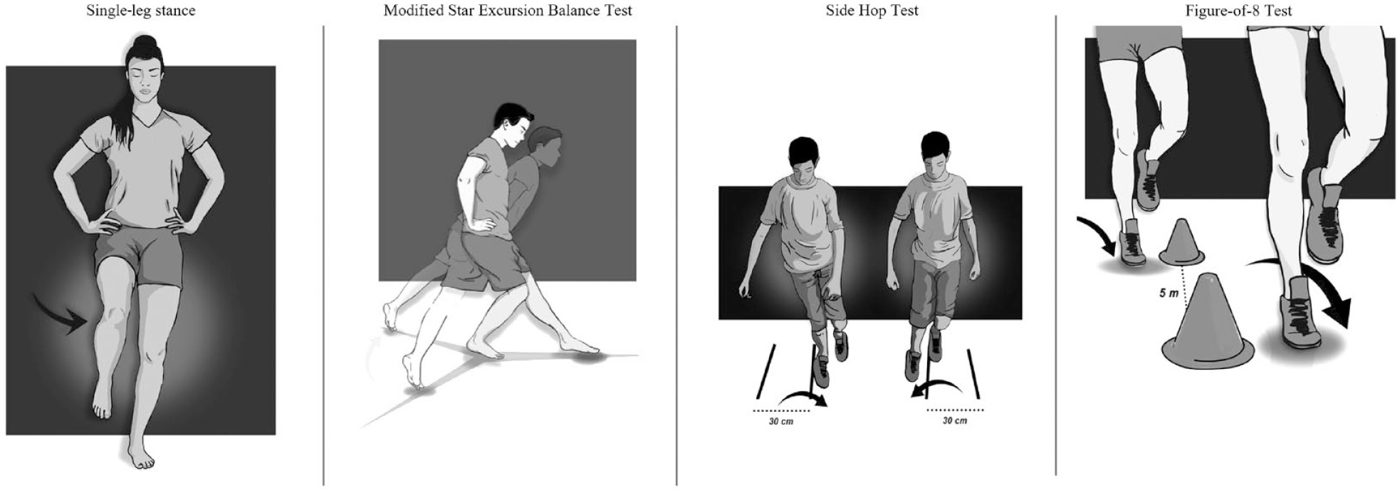
Ankle-GO 是把這些變成分數的工具:4 項功能測驗加 2 份問卷,滿分 25 分11。研究發現:
這些測驗要由物理治療師或防護員施測,結果再搭配臨床評估一起判斷。
專業補充:Ankle-GO 測驗方法、計分與研究數據
ROAST:急性期的臨床評估
國際踝關節聯盟(International Ankle Consortium)的共識8:急性外側踝扭傷的最低評估標準,包括確認受傷機轉,以及評估踝關節骨骼與韌帶。他們也提出 ROAST(以復健為導向的評估),用來找出與 CAI 有關的機械性與感覺動作缺損。
較早的回顧也建議用背屈弓箭步測試、SEBT、敏捷 T 測驗與垂直跳等功能測驗,搭配心理層面的評估,判斷能否重返運動38。
PAASS 告訴我們「要看什麼」,但沒有指定測驗。Ankle-GO 就是依照 PAASS 等文獻挑出測驗,組成可以計分的工具11。
- 單腳站立(SLS):赤腳單腳站、膝微彎約 10°、雙手叉腰、閉眼 20 秒,計算錯誤次數(手離開髖、抬腳跟或前腳掌、睜眼等)11。
- 改良星狀偏移平衡測驗(mSEBT):站在 Y 字中央,對側腳往前、後內、後外三個方向盡量伸遠,距離以腿長標準化,三方向平均為綜合分數(COMP)11。
- 側向跳(SHT):在相距 30 公分的兩條線之間,盡快左右來回跳 10 次11。
- 8 字形測驗(F8T):繞著相距 5 公尺的兩個標誌,用單腳跳盡快完成兩圈(共 20 公尺)11。
- 兩份問卷:FAAM(日常生活與運動兩個分量表)與 ALR-RSI(重返運動的心理準備度)11。
四項功能測驗中,沒有出現不穩定感就各加 1 分。Hertel 等人 2006 年發現,SEBT 八個方向中後內方向最能代表整體表現14,這也是 Ankle-GO 對後內方向另外加分的原因之一。
計分方式
| 項目 | 結果與得分 | 不穩定感/加分 | 滿分 |
|---|---|---|---|
| SLS | 錯誤 >3 次:0|1–3 次:1|0 次:2 | 無不穩定感 +1 | 3 |
| mSEBT | COMP <90%:0|90–95%:2|>95%:4 | 前方 >60% +1、後內 >90% +1、無不穩定感 +1 | 7 |
| SHT | >13 秒:0|10–13 秒:2|<10 秒:4 | 無不穩定感 +1 | 5 |
| F8T | >18 秒:0|13–18 秒:1|<13 秒:2 | 無不穩定感 +1 | 3 |
| FAAM-ADL | <90%:0|90–95%:1|>95%:2 | — | 2 |
| FAAM-Sport | <80%:0|80–95%:1|>95%:2 | — | 2 |
| ALR-RSI | <55%:0|55–63%:1|63–76%:2|>76%:3 | — | 3 |
計分表依 Picot 等人 2023 年原始論文 Table 3 整理11。
研究怎麼說
| 研究 | 重點結果 |
|---|---|
| Picot 2023,64 位病人+30 位健康者11 | 傷後 2 個月 7.7 ± 4 分、4 個月 13.9 ± 4.6 分、健康者 19.6 ± 3.4 分。內部一致性 α = 0.79,再測信度 ICC = 0.99,最小可偵測變化 1.2 分。2 個月分數預測 4 個月回到傷前水準,AUC 0.77;低於 8 分較不可能回到傷前水準。 |
| Picot 2024,兩年追蹤12 | 2 個月時低於 8 分,兩年內再扭傷風險約高 9 倍(OR 8.6)。 |
| Picot 2024,一年追蹤15 | 2 個月時高於 11 分,一年後成為 coper(回到原運動、沒有功能下降與再扭傷)的機率較高(OR 12.1)。 |
Ankle-GO 互動計分器(給治療師、教練)
預設值是一位虛構的範例個案。研究的預測數據都以「傷後 2 個月」的分數為準。
落在級距邊界上的數值(例如剛好 90% 或 13 秒)歸入中間級距。這個計分器只用來對照研究結果,不能取代臨床判斷;作者團隊另有官方的 Ankle-GO 應用程式11。
專業補充:PAASS 與 Ankle-GO 怎麼對照
以下依兩篇原始論文整理的對照,原作者沒有提出這張表:
| PAASS 領域 | Ankle-GO 對應項目 |
|---|---|
| 感覺動作控制 | SLS、mSEBT |
| 運動功能表現 | SHT、F8T |
| 踝關節缺損、運動員感受 | FAAM、測驗中的不穩定感 |
| 心理準備度 | ALR-RSI |
| 疼痛 | Ankle-GO 沒有納入,需另外評估 |
10. 怎麼預防再扭到?
整合結論
- 扭傷後能走路,不代表恢復完成;約四成會變成一扭再扭1。
- 剛扭到先保護、及早開始有人指導的運動;消炎止痛藥使用要謹慎2。
- 一扭再扭的人,要組合平衡、肌力與徒手治療5,並至少練 4 週以上23。
- 回場上要看客觀測驗,例如 Ankle-GO:傷後 2 個月低於 8 分,要再加強復健11。
補充:Ottawa 足踝準則
Ottawa 足踝準則(Ottawa Ankle Rules)用來判斷急性踝扭傷後是否需要照 X 光排除骨折41:
- 照踝關節 X 光:踝部疼痛,且外踝或內踝的遠端 6 公分的後緣或踝尖有骨頭壓痛,或受傷當下和就診時都無法走 4 步。
- 照足部 X 光:中足疼痛,且舟狀骨或第五蹠骨基部有骨頭壓痛,或受傷當下和就診時都無法走 4 步。
原始驗證研究中,準則對踝部與中足骨折的敏感度都是 100%,估計可減少約三分之一的 X 光檢查39。統合 27 篇研究、15,581 位病人的系統性回顧也顯示敏感度接近 100%,但特異度普通,可減少 30% 到 40% 不必要的 X 光40。也就是說,準則陰性時可以放心排除骨折;準則陽性不代表一定骨折。
補充:星狀偏移平衡測驗(SEBT)
星狀偏移平衡測驗(Star Excursion Balance Test, SEBT)是評估動態平衡的功能測驗:受測者單腳站在地上 8 條線(彼此相隔 45 度)的中央,另一腳盡量往各方向伸遠、輕觸地面後回到中央,記錄伸展距離29。
- 可以用來做什麼:系統性回顧認為 SEBT 信度良好,可以找出下肢傷害後的動態平衡缺損、預測下肢受傷風險,對訓練介入的效果也有反應42。
- 不必每次測 8 個方向:8 個方向之間重複性很高,後內方向最能代表整體表現;CAI 病人在前內、內側、後內三個方向的缺損最明顯14。
- 改良版(mSEBT)只測三個方向:前方、後內、後外。距離除以腿長(髂前上棘到內踝)標準化,三方向平均即為綜合分數(COMP);Ankle-GO 是先練習 4 次、再記錄 3 次取平均11。
分數怎麼看
- 受傷風險:235 位高中籃球員中,左右腳前方伸展差距大於 4 公分,下肢受傷機率約高 2.5 倍;女生綜合分數低於腿長的 94%,受傷機率約高 6.5 倍43。
- Ankle-GO 的計分門檻:綜合分數 90% 與 95% 為分級點,前方大於 60%、後內大於 90% 各加 1 分11。
SEBT 和 Y Balance 可以混用嗎?
不建議。20 位健康男性分別做 SEBT 和 Y Balance Test:前方方向在 SEBT 上伸得比較遠,後內與後外則沒有差異44。追蹤同一個人時,前後要用同一種測法,門檻值也要對應原研究使用的版本。
參考文獻(44 篇)點開查看
- Picot B, et al. Targeting visual-sensory and cognitive impairments following lateral ankle sprains, Part 1. Front Sports Act Living. 2025. DOI
- Vuurberg G, et al. Diagnosis, treatment and prevention of ankle sprains: update of an evidence-based clinical guideline. Br J Sports Med. 2018. DOI
- Dubois B, Esculier JF. Soft-tissue injuries simply need PEACE and LOVE. Br J Sports Med. 2020;54(2):72-73. DOI
- Chen, et al. MRI-based predictors and risk constellations of chronic ankle instability after acute lateral ankle sprain: a multicenter study. Acad Radiol. 2026. DOI
- Yang, et al. Treatment effects of physical therapy approaches to managing chronic ankle instability: a network meta-analysis of randomized controlled trials. J Orthop Sports Phys Ther. 2025. DOI
- Hou, et al. Efficacy of hip strengthening on balance and function outcomes in chronic ankle instability: a systematic review and meta-analysis. BMC Musculoskelet Disord. 2026. DOI
- Yin, et al. Effect of proprioceptive neuromuscular facilitation on patients with chronic ankle instability: a systematic review and meta-analysis. PLoS One. 2025. DOI
- Delahunt E, et al. Clinical assessment of acute lateral ankle sprain injuries (ROAST): 2019 consensus statement and recommendations of the International Ankle Consortium. Br J Sports Med. 2018. DOI
- Li Y, et al. Return to sport after anatomic lateral ankle stabilization surgery for chronic ankle instability: a systematic review and meta-analysis. Am J Sports Med. 2024;52(2):555-566. DOI
- Vopat ML, et al. Early versus delayed mobilization postoperative protocols for lateral ankle ligament repair: a systematic review and meta-analysis. Orthop J Sports Med. 2020. DOI
- Picot B, et al. Development and validation of the Ankle-GO score for discriminating and predicting return-to-sport outcomes after lateral ankle sprain. Sports Health. 2023(2024;16(1):47-57). DOI
- Picot B, et al. Low Ankle-GO score while returning to sport after lateral ankle sprain leads to a 9-fold increased risk of recurrence: a two-year prospective cohort study. Sports Med Open. 2024;10:23. DOI
- Smith MD, et al. Return to sport decisions after an acute lateral ankle sprain injury: introducing the PAASS framework. Br J Sports Med. 2021. DOI
- Hertel J, et al. Simplifying the star excursion balance test: analyses of subjects with and without chronic ankle instability. J Orthop Sports Phys Ther. 2006;36(3):131-7. DOI
- Picot B, et al. Ankle-GO score is associated with the probability of becoming coper after lateral ankle sprain: a 1-year prospective cohort study. Br J Sports Med. 2024;58(19):1115-1122. DOI
- Wu V, Padilla CA, Smith NA. Management of acute ankle sprains: common questions and answers. Am Fam Physician. 2025;112(6):609-617.(PubMed 41533404)
- Halder P, et al. Optimizing soft tissue injury rehabilitation: PEACE & LOVE with vs without cryotherapy — protocol for a sham-controlled randomized trial. Trials. 2026. DOI
- Pourkazemi F, et al. Predictors of chronic ankle instability after an index lateral ankle sprain: a systematic review. J Sci Med Sport. 2014;17(6):568-73. DOI
- Wijnhoud EJ, et al. One in three patients with chronic lateral ankle instability has a cartilage lesion. Am J Sports Med. 2023;51(7):1943-1951. DOI
- Anderson DD, et al. Ankle osteoarthritis: toward new understanding and opportunities for prevention and intervention. J Orthop Res. 2024;42(12):2613-2622. DOI
- Owoeye OBA, et al. Health-related outcomes 3-15 years following ankle sprain injury in youth sport. Foot Ankle Int. 2022;43(1):21-31. DOI
- Bott KN, et al. Subchondral bone degeneration and pathology 3-15 years following ankle sprain injury in adolescent sport. Foot Ankle Int. 2025;46(1):19-28. DOI
- Zhang C, et al. Effectiveness of exercise therapy on chronic ankle instability: a meta-analysis. Sci Rep. 2025;15:11709. DOI
- Faghihi R, Khanmohammadi R. Comparing virtual reality and balance training effects on postural strategies during ball kicking in soccer players with chronic ankle instability. Sci Rep. 2024. DOI
- Sarkhosh SS, Khanmohammadi R. Comparison of kinetic adaptations in gait initiation following exergaming and balance training in athletes with chronic ankle instability. Sci Rep. 2026. DOI
- Wang B, et al. A randomized controlled trial comparing rehabilitation with isokinetic exercises and Thera-Band strength training in patients with functional ankle instability. PLoS One. 2022;17(12):e0278284. DOI
- Hall EA, et al. Strength-training protocols to improve deficits in participants with chronic ankle instability: a randomized controlled trial. J Athl Train. 2015;50(1):36-44. DOI
- Liao W, et al. The interventional effects of PNF training on functional ankle instability in habitually active adults. Front Public Health. 2026. DOI
- Alahmari KA, et al. Effectiveness of low-frequency stimulation in proprioceptive neuromuscular facilitation techniques for post ankle sprain balance and proprioception in adults: a randomized controlled trial. Biomed Res Int. 2020:9012930. DOI
- Kong DH, et al. Effectiveness of hospital-based systemic rehabilitation in improving ankle function after surgery in chronic ankle instability patients. Biomed Res Int. 2021:6695096. DOI
- Zhao Y, et al. Functional outcomes of immediate weightbearing after arthroscopic lateral ankle ligament repair: a prospective randomized single-center trial. Am J Sports Med. 2024;52(14):3618-3624. DOI
- Miyamoto W, et al. Accelerated versus traditional rehabilitation after anterior talofibular ligament reconstruction for chronic lateral instability of the ankle in athletes. Am J Sports Med. 2014;42(6):1441-7. DOI
- Lee JH, et al. Proprioception and neuromuscular control at return to sport after ankle surgery with the modified Broström procedure. Sci Rep. 2022;12:610. DOI
- Du M, et al. Distal insertion rupture of lateral ankle ligament as a predictor of weakened and delayed sports recovery after acute ligament repair. BMC Musculoskelet Disord. 2022;23:294. DOI
- Luthfi APWY, et al. Risk factors for the recurrence of instability after lateral ankle ligament repair. Foot Ankle Int. 2023;44(7):617-628. DOI
- Zendeli F, et al. A smaller tibiotalar sector is a risk factor for recurrent anterolateral ankle instability after a modified Broström-Gould procedure. Foot Ankle Int. 2024;45(4):338-347. DOI
- Dallman J, et al. Current definitions of failure in lateral ankle instability surgery: a systematic review. Am J Sports Med. 2023;51(10):2748-2757. DOI
- Clanton TO, et al. Return to play in athletes following ankle injuries. Sports Health. 2012;4(6):471-4. DOI
- Stiell IG, et al. Decision rules for the use of radiography in acute ankle injuries: refinement and prospective validation. JAMA. 1993;269(9):1127-32. DOI
- Bachmann LM, et al. Accuracy of Ottawa ankle rules to exclude fractures of the ankle and mid-foot: systematic review. BMJ. 2003;326(7386):417. DOI
- Ottawa Ankle Rule(準則條文). MDCalc. 連結
- Gribble PA, Hertel J, Plisky P. Using the Star Excursion Balance Test to assess dynamic postural-control deficits and outcomes in lower extremity injury: a literature and systematic review. J Athl Train. 2012;47(3):339-57. DOI
- Plisky PJ, et al. Star Excursion Balance Test as a predictor of lower extremity injury in high school basketball players. J Orthop Sports Phys Ther. 2006;36(12):911-9. DOI
- Coughlan GF, et al. A comparison between performance on selected directions of the star excursion balance test and the Y balance test. J Athl Train. 2012;47(4):366-71. DOI
文獻資料經 PubMed 檢索核對。本文為衛教整理,不能取代個別的醫療評估。